Trauma1's Basic Interpretation Of Blood Work: Series #1 - Hepatic/Biliary Function!

Trauma1

Trauma1

Legend
Awards
3
  • RockStar
  • Legend!
  • Established
I'm putting together a thread here to help you all understand the basics of interpreting blood work. I plan to have a series of these threads, but i want to focus initially on Hepatic/Biliary function. As you all know, these blood work factors are important to monitor while on cycle, or if you have some concurrent medical issue. I'll answer any questions about bloodwork and such that you may have to the best of my abilities. :)


Steroids with Michael Scally, MD
Oral Anabolic Steroids, Liver Enzyme Tests and Liver Function
by Michael C. Scally, M.D.Author of eBook Human Experimentation in Anabolic Steroid Research by Michael Scally, M.D.
Harvard Medical School - M.D.; Harvard-M.I.T. Program In Health Science & Technology
Massachusetts Institute of Technology, B.S. Chemistry/LIfe Sciences


Dr. Scally early on recognized the lack of research and treatment for individuals using anabolic-androgenic steroids (AAS). He has remained as the sole physician by reputation and publication to actively pursue and advocate the proper use of AAS to optimize health. Dr. Scally has personally cared for thousands of individuals using AAS. His protocol for Anabolic Steroid Induced Hypogonadism has been presented before the Endocrine Society, American Association of Clinical Endocrinologists, American College of Sports Medicine, & International Workshop on Adverse Drug Reactions and Lipodystrophy in HIV.


Question:


Do oral steroids have long-term effects on liver function long after they have been discontinued? I have done quite a few cycles of anadrol and dianabol in the past. But I haven’t done any oral AAS, prohormones, legal or otherwise in several years and my liver function tests are still elevated (AST and ALT). They are about double the top of the normal range. Can any other factors account for this e.g. dietary supplements, genetics, intense physical exercise, heavy childhood use of NSAIDs?


Response:


Mild elevations in liver chemistry tests such as alanine aminotransferase (ALT) and aspartate aminotransferase (AST) can reveal serious underlying conditions or have transient and benign etiologies. There are no controlled clinical trials examining the optimal approach for evaluating serum liver chemistries. The American Gastroenterological Association guideline regarding the evaluation and management of abnormal liver chemistry tests proposes a practical, algorithmic approach when the history and physical examination do not reveal the cause.

The history should be thorough, with special attention given to the use of medications, vitamins, herbs, drugs, and alcohol; family history; and any history of blood-product transfusions.[1] In addition to liver chemistries, an initial serologic evaluation includes a prothrombin time; albumin; complete blood count with platelets; hepatitis A, B, and C serologies; and iron studies. The most common causes of elevated aminotransferase levels include alcohol-related liver injury, chronic hepatitis B and C, autoimmune hepatitis, hepatic steatosis (fatty infiltration of the liver), nonalcoholic steatohepatitis, hemochromatosis, Wilson's disease, alpha1-antitrypsin deficiency, and celiac sprue.

Depending on the etiology, management strategies may include cessation of alcohol use, attention to medications, control of diabetes, and modification of lifestyle factors such as obesity. If elevations persist after an appropriate period of observation, further testing may include ultrasonography, other serum studies, and in some cases, liver biopsy.[2] Isolated alterations of biochemical markers of liver damage in a seemingly healthy patient often represent a challenge even for the experienced clinician and usually set off a battery of further, costly tests and consultations that may ultimately prove unnecessary.

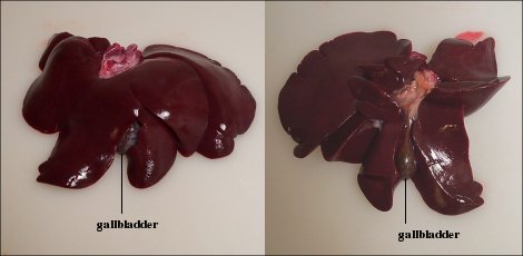
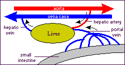
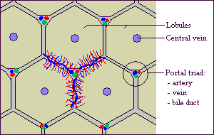
The liver is the largest and most metabolically complex organ in humans. The liver receives a dual blood supply. The portal vein drains the splanchnic, viscera, circulation and provides 75% of the total blood flow. The hepatic artery provides the remaining 25%. The hepatic vein carries all efferent blood to the inferior vena cava. Rich supplies of lymphatic vessels also drain the liver.

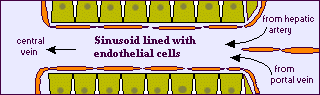
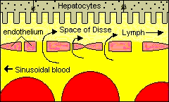
The liver is a complex organ with interdependent metabolic, excretory, and defense functions. Hepatocytes make up the bulk of the organ. Sinusoidal lining cells comprise at least four distinct populations: endothelial cells, Kupffer's cells, perisinusoidal fat-storing cells and pit cells. Endothelial cells are responsible for endocytosis of molecules and particles, and play a role in lipoprotein metabolism. Spindle-shaped Kupffer's cells are tissue macrophages. Perisinusoidal fat-storing cells (Ito cells) store vitamin A. Pit cells are large, granular lymphocytes, which function as natural killer cells.

The liver plays a central role in carbohydrate, protein, and fat metabolism. It stabilizes glucose level by taking up and storing glucose as glycogen (glycogenesis), breaking glycogen down to glucose (glycogenolysis), and forming glucose from noncarbohydrate sources (gluconeogenesis). The liver synthesizes the majority of proteins that circulate in the plasma, including albumin and most of the globulins other than gamma globulins. It is responsible for synthesizing and secreting bile and plasma proteins, including clotting factors. The liver is the site of most amino acid interconversions and catabolism. Amino acid deamination produces urea and esterification of fatty acids produces triglycerides. The liver packages triglycerides with cholesterol, phospholipids, and an apoprotein into a lipoprotein. The lipoprotein enters blood for utilization or storage in adipocytes. Most cholesterol synthesis takes place in the liver.

The liver detoxifies noxious substances arriving from the splanchnic (viscera) circulation, preventing them from entering the systemic circulation. This particularly makes the liver susceptible to drug-induced injury. The liver converts some lipophilic compounds into more water-soluble agents and others to less active agents. In conjunction with the spleen, it is involved in the destruction and reclamation of spent red blood cells.

Prior to a discussion of liver pathology, it is important to have an understanding in the interpretation of laboratory tests. Normal refers to a theoretical frequency distribution for a set of variable data, usually represented by a bell-shaped curve symmetrical about the mean. Laboratory values for a reference range are from a group of healthy individuals with no known factors (medications, illness, genetics, etc.) that would influence the outcome of the testing. The reference range for a particular laboratory test is dependent upon a given subpopulation (e.g., male, female, or children) and the testing laboratory or manufacturer. Federal regulations require laboratories to adhere to certain standards. "Prior to reporting patient test results, the laboratory must verify or establish, for each method, the performance specifications for the following performance characteristics: accuracy; precision; analytical sensitivity and specificity, if applicable; the reportable range of patient test results; the reference range(s) (normal values); and any other applicable performance characteristic."[3] The normal reference range typically refers to the mean or average +2 standard deviations.[4] Interpretation of results is being either within, normal, for a value falling within this bell-shaped curve (reference range) or outside, abnormal, the reference range. Accordingly, 2.5% of normal patients have "abnormal" aminotransferase levels.

A basic tenet, standard practice, of medicine is that interpretation of results is within the framework of a patient's medical condition and treatment, the overall health of the patient.[5] Physicians are taught to think about clinical testing in terms of the clinical significance (particularly, predictive value) of a given test in a given situation. All tests have strengths and limitations for their use in reaching a certain diagnosis or making a causal inference. The risk of a test is seldom inherent in the test itself, but rather is a function of the context in which use of the test is providing information for medical decision-making. Many factors affect test results including sex, medications, overall health of the individual, temporal influences, and variations in laboratory techniques. Thus, in terms of diagnosis, interpretation of a diagnostic test is in the context of history, examination, other tests, and other relevant medical considerations.[6] The proper and correct interpretation for a test is within the situational context.

Levels of serum liver enzymes are indications of hepatocyte integrity or cholestasis rather than liver function. A change in serum protein levels or clotting times may be associated with a decrease in liver functioning mass, although neither is specific for liver disease. No single or simple test assesses overall liver pathology. Use of several screening tests improves the detection of hepatobiliary abnormalities, differentiates the basis for clinically suspected disease, and determines the severity of liver disease (hepatocytes (hepatocellular dysfunction), the biliary excretory apparatus (cholestasis), and the vascular system (portal hypertension)).

The widespread availability and use of serum blood chemistries for screening both symptomatic and asymptomatic patients has resulted in a dramatic increase in the number of normal and abnormal liver chemistry tests requiring interpretation by physicians. A number of review articles on the evaluation of abnormal liver function tests are available on the internet.[7] Aminotransferases (transaminase) include alanine aminotransferase (ALT) and aspartate aminotransferase (AST). Both are exquisitely sensitive indicators of hepatocellular injury and provide the best guide to hepatocellular necrosis/inflammation.[8]

ALT (8-37 IU/L) is present in hepatocytes (liver cells) and is reliable for routine screening for liver disease. It is also called serum glutamate pyruvate transaminase (SGPT) or alanine aminotransferase (ALAT). When a cell is damaged, it leaks this enzyme into the blood, where it is measured. ALT rises dramatically in acute liver damage, such as viral hepatitis or paracetamol (acetaminophen) overdose. The highest level of ALT is in the liver, and levels of this enzyme are accordingly more specific indicators of liver injury. The magnitude of the elevation has no prognostic value and does not correlate with the degree of liver damage.

AST (10-34 IU/L), also called serum glutamic oxaloacetic transaminase (SGOT) or aspartate aminotransferase (ASAT/AAT) is similar to alanine transaminase (ALT) in that it is another enzyme associated with liver parenchymal cells. AST is present, in decreasing order of concentration, in the liver, cardiac muscle, skeletal muscle, kidneys, brain, pancreas, lungs, leukocytes, and erythrocytes. AST levels thus rise in MI, heart failure, muscle injury, CNS disease, and other nonhepatic disorders. AST is relatively nonspecific, but high levels indicate liver cell injury. In most liver diseases, the AST increase is less than that of ALT (AST/ALT ratio < 1).

Both aminotransferases are normally present in serum at low levels, usually less than 30 to 40 IU/L. The normal range varies widely among laboratories. The following table lists factors affecting AST and ALT serum activity, other than liver injury.[9] Release of both enzymes into the blood occurs in increasing amounts with liver cell membrane damage. Necrosis of liver cells is not required for the release of the aminotransferases. In fact, there is poor correlation between the degree of liver-cell damage and the level of the aminotransferases. The magnitude of elevation covers a very wide range. Levels <100 IU are common and nonspecific, and often have no clinical significance; levels of 100-300 IU are seen in numerous mild/moderate inflammatory processes. In acute viral or drug hepatitis aminotransferase levels are typically in the 500-1,500 IU range, but in alcoholic hepatitis they are usually <300 IU, even if the disease is severe. Values >3,000 IU usually are seen only in acute toxic necrosis or severe hypoxia ("shock liver," "ischemic hepatitis"); in both disorders levels typically plummet within two to three days, whereas values fall more slowly in viral hepatitis. Aminotransferase levels are variable in biliary obstruction but usually remain <200 IU, except with acute passage of common duct stone, characterized by a sudden rise to hepatitic levels and a rapid fall over the next one to two days.


Factor
AST
ALT
Comments​


Factor: Time of day

AST: 45% variation during day; highest in afternoon, lowest at night

ALT: No significant difference between 0900 and 2100;

Comment: similar in liver disease and health


Factor: Day-to-day

AST: 5–10% variation from one day to next

ALT: 10–30% variation from one day to next

Comment: Similar in liver disease and health, and in elderly and young


Factor: Race/gender

AST: 15% higher in African-American men

ALT: No significant difference between African-American, other women


Factor: BMI (body mass index)

AST: 40–50% higher with high BMI

ALT: 40–50% higher with high BMI

Comment: Direct relationship between weight and AST, ALT


Factor: Meals

AST: No effect

ALT: No effect


Factor: Exercise

AST: Threefold increase with strenuous exercise
20% lower in those who exercise at usual levels than in those who do not exercise or exercise more strenuously than usual

ALT: Effect of exercise seen predominantly in men; minimal difference in women (<10%). Enzymes increase more with strength training


Factor: Specimen storage

AST: Stable at room temp for 3 days, in refrigerator for 3 weeks (<10% decrease); stable for years frozen (10–15% decrease)

ALT: Stable at room temperature for 3 days, in refrigerator for 3 weeks (10–15% decrease); marked decrease with freezing/thawing

Comment: Stability based on serum separated from cells; stable for 24 h in whole blood, marked increase after 24 h


Factor: Hemolysis, hemolytic anemia:

AST: Significant increase

ALT: Moderate increase attributable to release from red cell

Comment: Dependent on degree of hemolysis; usually several fold lower than increases in lactate dehydrogenase (LDH)


Factor: Muscle injury:

AST: Significant increase

ALT: Moderate increase

Comment: Related to amount of increase in creatine kinase (CK)


Other biochemical tests of interest are γ-glutamyl transpeptidase (GGT), lactic dehydrogenase (LDH), alkaline phosphatase (ALP), albumin, and bilirubin. Corresponding changes in the serum levels of these markers assist in defining the etiology. γ-Glutamyl transpeptidase (GGT), also known as γ-glutamyltransferase, is present in the liver, pancreas, and kidney. GGT transfers the γ-glutamyl group from one peptide to another or to an L-amino acid. GGT levels (0-51 IU/L) are elevated in diseases of the liver, biliary tract, and pancreas with obstruction of the common bile duct. Drug use and alcohol (acute and chronic) ingestion also elevate GGT. GGT may be elevated with even minor, sub-clinical levels of liver dysfunction. Alkaline phosphatase (ALP) is an enzyme in the cells lining the biliary ducts of the liver. ALP levels (44-147 IU/L) in plasma will rise with large bile duct obstruction, intrahepatic cholestasis, or infiltrative diseases of the liver. ALP is also present in bone. Serum γ-glutamyl transpeptidase (GGT) activity correlates closely with the activities of alkaline phosphatase (ALP) in various forms of liver disease. Maximum elevations of the enzyme activities are observed in diseases that affect the biliary tract. Compared with ALP, GGT is generally increased to a greater extent and is thus the most sensitive indicator of biliary-tract disease.

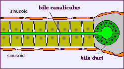
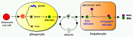
Lactic dehydrogenase (LDH) is commonly included in routine analysis, is insensitive as an indicator of hepatocellular injury, but is better as a marker for hemolysis, myocardial infarction (heart attack), or pulmonary embolism. LDH can be quite high with malignancies involving the liver. Albumin (3.9-5.0 g/dL) is a protein made specifically by the liver, and can be measured cheaply and easily. It is the main constituent of total protein; the remaining fraction is called globulin (including the immunoglobulins). Bilirubin is a breakdown product of heme (a part of hemoglobin in red blood cells). The liver is responsible for clearing the blood of bilirubin. Bilirubin is taken up into hepatocytes, conjugated (modified to make it water-soluble), and secreted into the bile, which is excreted into the intestine. Increased total bilirubin causes jaundice, and can signal a number of problems.

Elevated serum aminotransferase levels, especially aspartate aminotransferase levels, may be caused by disorders that affect organs or tissues other than the liver, with the most common being striated muscle. Conditions or activities that can cause such elevations include subclinical inborn errors of muscle metabolism; acquired muscle disorders, such as polymyositis; and exercise. If striated muscle is the source of increased aminotransferase levels, serum levels of creatine kinase will be elevated to the same degree or to an even higher degree.

Creatine kinase (CK), also known as phosphocreatine kinase or creatine phosphokinase (CPK) is an enzyme that catalyses the conversion of creatine to phosphocreatine. In tissues that consume ATP rapidly, especially skeletal muscle, but also brain and smooth muscle, phosphocreatine serves as an energy reservoir for the rapid regeneration of ATP. Clinically, creatine kinase is assayed in blood tests as a marker of myocardial infarction (heart attack), rhabdomyolysis (muscle breakdown), and in acute renal failure. Numerous studies have evaluated changes in CK activity after exercise and found that it differs markedly according to exercise conditions. In isometric muscle contraction exercise, peak serum CK activity is observed relatively early, 24-48 hours after exercise, whereas it is seen 3-7 days after exercise in eccentric muscle contraction exercise, and a biphasic pattern is observed in weight training.

Toxic effects of AAS on the liver are primarily due to 17α-alkylated steroids and reported to include increased enzyme activities, cholestasis, peliosis hepatis adenoma, and even case reports of carcinoma.[10] The use of anabolic steroids is common among athletes, particularly bodybuilders. Prior reports of anabolic steroid-induced hepatotoxicity based on elevated aminotransferase levels have been overstated. Such reports may have misled the medical community to emphasize steroid-induced hepatotoxicity when interpreting elevated aminotransferase levels and disregard muscle damage. Levels of both aspartate aminotransferase (AST) and alanine aminotransferase (ALT) may increase with strenuous exercise. Evaluating enzyme elevations in patients who use anabolic steroids, physicians should consider the CK and GGT levels as essential elements in distinguishing muscle damage from liver damage
 
Trauma1

Trauma1

Legend
Awards
3
  • RockStar
  • Legend!
  • Established
UTH Gastroenterology Grand Rounds - Discussion


--------------------------------------------------------------------------------

Final Diagnosis: Drug Induced Cholestasis
Drug induced cholestasis is a well-known side effect of many performance-enhancing supplements. Often times, these are used discretely by athletes and body builders. Steroids are believed to increase muscle mass by binding to androgen receptors and increasing protein synthesis. It also has an anticatabolic effect in muscle. Gynecomastia occurs from the peripheral conversion of androgens to estradiol (1). (Visual 2) Testosterone first appeared in international sporting competitions after the 1948 Olympics. East German records reported a 0.7 second reduction in a 100m sprint, 7-10 seconds in the 1500m run, 8-15m in a javelin throw (2). Names of common anabolic steroids are oxymetholone, stanozolol, nandrolone, and testosterone.


Liver Injury

Liver injury is an increase of ALT or conjugated bilirubin greater than two times the upper limit of normal or a combined increase of AST, alkaline phosphatase, total bilirubin and at least one parameter is greater than two times the upper limit of normal. The AASLD conference in 2000 suggested that an ALT of more than three times the upper limit of normal and a total bilirubin of more than twice the upper limit of normal be used to indicate significant abnormalities on the liver test (3).


Liver injury is broadly classified into

1) hepatocellular injury with increases in ALT

2) cholestatic with an elevated alkaline phosphatase

3) mixed with elevated alkaline phosphatase and ALT.


Pathogenesis

The cause of the liver injury and subsequent elevations of liver enzymes are drug specific. The various mechanisms include the inhibitions of drug metabolism, disruption of bilirubin transport pumps causing cholestasis, apoptosis via Fas pathways, free radiation formation, and mitochondrial dysfunction. Anabolic steroid –induced jaundice appears to be from interference of bilirubin excretion into the canaliculus via suppression of the Na-K adenosine triphosphatase activity (6). The cholestatic side effects are typically seen in C17 alpha alkylated steroids.


Clinical presentation/Diagnosis

Anabolic steroid induced cholestasis typically causes pruritis and jaundice as an initial presenting symptom with elevations in the alkaline phosphatase and bilirubin level. In rare cases, chronic liver injury may lead to the vanishing bile duct syndrome (3). There has been case reports of marked elevation of AST/ALT from steroid use (4).

Acute hepatotoxicity is associated with malaise, pain, and jaundice. Labs will show an elevation of ALT and a mild increase in alkaline phosphatase levels. Increased PT/INR and encephalopathy may also be seen in patients with liver failure.

Work up for a drug induced hepatotoxicity includes excluding other precipitating factors (viral, biliary obstruction, alcohol abuse, autoimmune causes, Wilson’s, Hematochromatosis, shock liver, etc). Biliary imaging is important to evaluate for biliary obstruction. A review of the medications should be done to evaluate for potential hepatotoxic medications. (Visual 3)

When urine is tested for androgen use, the ration of testosterone and epitestosterone (inactive metabolite of testosterone produced by the gonads with little peripheral metabolism of testosterone) is measured. The normal ratio is 1:1. If exogenous testosterone is used, the testosterone:epitestosterone is increased (1).


Management

The suspected drug or drugs should be stopped. In most cases, the liver parameters will improve after stopping the offending agent, however the improvement may be delayed or follow a protracted course over the next few weeks/months. Liver dysfunction may also briefly worsen following discontinuation of the offending medication. Cholestyramine or ursodiol may provide symptomatic relief.


Patient Follow-up

Patient was diagnosed with drug-induced cholestasis. He was using anabolic steroids and tamoxifen which commonly causes a cholestatic liver disease. Tamoxifen is used by body builders for its anti-estrogen effects to reduce anabolic steroid-induced gynecomastia. The patient was instructed to stop all performance enhancing drugs and start cholestyramine and ursodiol. His jaundice and pruritis gradually improved over the next 2 months.
 
Trauma1

Trauma1

Legend
Awards
3
  • RockStar
  • Legend!
  • Established
Liver Function Tests:


Liver function tests represent a broad range of normal functions performed by the liver. The diagnosis of liver disease depends upon a complete history, complete physical examination, and evaluation of liver function tests and further invasive and noninvasive tests. Many patients become confused regarding the meaning of a liver function test. This section is designed to describe the basic liver function tests and the meaning for patients.

The hepatobiliary tree represents hepatic cells and biliary tract cells. Inflammation of the hepatic cells results in elevation in the alanine aminotransferase (ALT), aspartate aminotransferase (AST) and possibly the bilirubin. Inflammation of the biliary tract cells results predominantly in an elevation of the alkaline phosphatase. In liver disease there are crossovers between purely biliary disease and hepatocellular disease. To interpret these, the physician will look at the entire picture of the hepatocellular disease and biliary tract disease to determine which is the primary abnormality.

Alanine Aminotransferase (ALT):

ALT is the enzyme produced within the cells of the liver. The level of ALT abnormality is increased in conditions where cells of the liver have been inflamed or undergone cell death. As the cells are damaged, the ALT leaks into the bloodstream leading to a rise in the serum levels. Any form of hepatic cell damage can result in an elevation in the ALT. The ALT level may or may not correlate with the degree of cell death or inflammation. ALT is the most sensitive marker for liver cell damage.

Aspartate Aminotransferase (AST):

This enzyme also reflects damage to the hepatic cell. It is less specific for liver disease. It may be elevated and other conditions such as a myocardial infarct (heart attack). Although AST is not a specific for liver as the ALT, ratios between ALT and AST are useful to physicians in assessing the etiology of liver enzyme abnormalities.

Alkaline Phosphatase:

Alkaline phosphatase is an enzyme, which is associated with the biliary tract. It is not specific to the biliary tract. It is also found in bone and the placenta. Renal or intestinal damage can also cause the alkaline phosphatase to rise. If the alkaline phosphatase is elevated, biliary tract damage and inflammation should be considered. However, considering the above other etiologies must also be entertained. One way to assess the etiology of the alkaline phosphatase is to perform a serologic evaluation called isoenzymes. Another more common method to asses the etiology of the elevated alkaline phosphatase is to determine whether the GGT is elevated or whether other function tests are abnormal (such as bilirubin)

Alkaline phosphatase may be elevated in primary biliary cirrhosis, alcoholic hepatitis, PSC, gallstones in choledocholithiasis.

Gamma Glutamic Transpeptidase (GGT):

This enzyme is also produced by the bile ducts. However, it is not very specific to the liver or bile ducts. It is used often times to confirm that the alkaline phosphatase is of the hepatic etiology. Certain GGT levels, as an isolated finding, reflect rare forms of liver disease. Medications commonly cause GGT to be elevated. Liver toxins such as alcohol can cause increases in the GGT.

Bilirubin:

Bilirubin is a major breakdown product of hemoglobin. Hemoglobin is derived from red cells that have outlived their natural life and subsequently have been removed by the spleen. During splenic degradation of red blood cells, hemoglobin (the part of the red blood cell that carries oxygen to the tissues) is separated out from iron and cell membrane components. Hemoglobin is transferred to the liver where it undergoes further metabolism in a process called conjugation. Conjugation allows hemoglobin to become more water-soluble. The water solubility of bilirubin allows the bilirubin to be excreted into bile. Bile then is used to digest food.

As the liver becomes irritated, the total bilirubin may rise. It is then important to understand the difference between total bilirubin, which has undergone conjugation (that is hepatic cell metabolism), and at portion of bilirubin which has not been metabolized. These two components are called total bilirubin and direct bilirubin. The direct bilirubin fraction is that portion of bilirubin that has undergone metabolism by the liver. When this fraction is elevated, the cause of elevated bilirubin (hyperbilirubinemia) is usually outside the liver. These types of causes are typically gallstones. This type of abnormality is usually treated with surgery (such as a gallbladder removal or choleycystectomy).

If the direct bilirubin is low, while the total bilirubin is high, this reflects liver cell damage or bile duct damage within the liver itself.


Albumin:

Albumin is the major protein present within the blood. Albumin is synthesized by the liver. As such, it represents a major synthetic protein and is a marker for the ability of the liver to synthesize proteins. It is only one of many proteins that are synthesized by the liver. However, since it is easy to measure, it represents a reliable and inexpensive laboratory test for physicians to assess the degree of liver damage present in the in any particular patient. When the liver has been chronically damaged, the albumin may be low. This would indicate that the synthetic function of the liver has been markedly diminished. Such findings suggest a diagnosis of cirrhosis. Malnutrition can also cause low albumin (hypoalbuminemia) with no associated liver disease.

Prothrombin time (PT):

Another measure of hepatic synthetic function is the prothrombin time. Prothrombin time is affected by proteins synthesized by the liver. Particularly, these proteins are associated with the incorporation of vitamin K metabolites into a protein. This allows normal coagulation (clotting of blood). Thus, in patients who have prolonged prothrombin times, liver disease may be present. Since a prolonged PT is not a specific test for liver disease, confirmation of other abnormal liver tests is essential. This may include reviewing other liver function tests or radiology studies of the liver. Diseases such as malnutrition, in which decreased vitamin K ingestion is present, may result in a prolonged PT time. An indirect test of hepatic synthetic function includes administration of vitamin K (10mg) subcutaneously over three days. Several days later, the prothrombin time may be measured. If the prothrombin time becomes normal, then hepatic synthetic function is intact. This test does not indicate that there is no liver disease, but is suggestive that malnutrition may coexist with (or without) liver disease.

Platelet count:

Platelets are cells that form the primary mechanism in blood clots. They're also the smallest of blood cells. They derived from the bone marrow from the larger cells known as megakaryocytes. Individuals with liver disease develop a large spleen. As this process occurs platelets are trapped with in the sinusoids (small pathways within the spleen) of the spleen. While the trapping of platelets is a normal function for the spleen, in liver disease it becomes exaggerated because of the enlarged spleen (splenomegaly). Subsequently, the platelet count may become diminished.

Serum protein electrophoresis:

This is an evaluation of the types of proteins that are present with in a patient's serum. By using an electrophoretic gel, major proteins can be separated out. This results in four major types of proteins. These are 1) albumin, 2) alpha globulins, 3) beta globulins and 4) gammaglobulins. This test is useful for evaluation of patients who have abnormal liver function tests since it allows a direct quantification of multiple different serum proteins. If the gamma globulin fraction is elevated, autoimmune hepatitis may be present. In addition a deficiency in the alpha globulin fraction can result in the diagnosis, or a clinical clue, to A. alpha-1 antitrypsin deficiency. This is a simple blood test that is commonly performed by hepatologists
 
Trauma1

Trauma1

Legend
Awards
3
  • RockStar
  • Legend!
  • Established
I'd be happy to answer specific questions if you all have any. Keep in mind now that blood work is pretty much worthless without a baseline to compare to; not to mention it isn't worth much either unless it's completed in a clinical setting where the patient's history/physical and other diagnostic tests can all be correlated to support a practitoner's medical diagnosis. That is not the point of this thread, however.

I'm here to help you guys acquire a basic understanding of blood work; nothing more. :cool:
 
Trauma1

Trauma1

Legend
Awards
3
  • RockStar
  • Legend!
  • Established
The Liver: Introduction and Index
--------------------------------------------------------------------------------

The liver is the largest gland in the body and performs an astonishingly large number of tasks that impact all body systems. One consequence of this complexity is that hepatic disease has widespread effects on virtually all other organ systems. At the risk of losing sight of the forest by focusing on the trees, we will focus on three fundamental roles of the liver:

Vascular functions, including formation of lymph and the hepatic phagocytic system.
Metabolic achievements in control of synthesis and utilization of carbohydrates, lipids and proteins.
Secretory and excretory functions, particularly with respect to the synthesis of secretion of bile.
The latter is the only one of the three that directly affects digestion - the liver, through its biliary tract, secretes bile acids into the small intestine where they assume a critical role in the digestion and absorption of dietary lipids. However, understanding the vascular and metabolic functions of the liver is critical to appreciating the gland as a whole.


Architecture of the Liver and Biliary Tract

--------------------------------------------------------------------------------

The liver lies in the abdominal cavity, in contact with diaphragm. Its mass is divided into several lobes, the number and size of which vary among species. In most mammals, a greenish sac - the gallbladder - is seen attached to the liver and careful examination will reveal the common bile duct, which delivers bile from the liver and gallbladder into the duodenum. The image below is of a liver from a dog, and illustrates these concepts. The panel on the left shows that aspect of the liver that faces the contents of the abdominal cavity. The right panel shows the flatter face of the liver which is in contact with the diaphragm.


Understanding function and dysfunction of the liver, more than most other organs, depends on understanding its structure. The major aspects of hepatic structure that require detailed attention include:

The hepatic vascular system, which has several unique characteristics relative to other organs
The biliary tree, which is a system of ducts that transports bile out of the liver into the small intestine
The three dimensional arrangements of the liver cells, or hepatocytes and their association with the vascular and biliary systems.
The Hepatic Vascular System
The circulatory system of the liver is unlike that seen in any other organ. Of great importance is the fact that a majority of the liver's blood supply is venous blood. The pattern of blood flow in the liver can be summarized as follows:

Roughly 75% of the blood entering the liver is venous blood from the portal vein. Importantly, all of the venous blood returning from the small intestine, stomach, pancreas and spleen converges into the portal vein. One consequence of this is that the liver gets "first pickings" of everything absorbed in the small intestine, which, as we will see, is where virtually all nutrients are absorbed.
The remaining 25% of the blood supply to the liver is arterial blood from the hepatic artery.


Terminal branches of the hepatic portal vein and hepatic artery empty together and mix as they enter sinusoids in the liver. Sinusoids are distensible vascular channels lined with highly fenestrated or "holey" endothelial cells and bounded circumferentially by hepatocytes. As blood flows through the sinusoids, a considerable amount of plasma is filtered into the space between endothelium and hepatocytes (the "space of Disse"), providing a major fraction of the body's lymph.
Blood flows through the sinusoids and empties into the central vein of each lobule.


Central veins coalesce into hepatic veins, which leave the liver and empty into the vena cava.
The Biliary System
The biliary system is a series of channels and ducts that conveys bile - a secretory and excretory product of hepatocytes - from the liver into the lumen of the small intestine. Hepatocytes are arranged in "plates" with their apical surfaces facing and surrounding the sinusoids. The basal faces of adjoining hepatocytes are welded together by junctional complexes to form canaliculi, the first channel in the biliary system. A bile canaliculus is not a duct, but rather, the dilated intercellular space between adjacent hepatocytes.

Hepatocytes secrete bile into the canaliculi, and those secretions flow parallel to the sinusoids, but in the opposite direction that blood flows. At the ends of the canaliculi, bile flows into bile ducts, which are true ducts lined with epithelial cells. Bile ducts thus begin in very close proximity to the terminal branches of the portal vein and hepatic artery, and this group of structures is an easily recognized and important landmark seen in histologic sections of liver - the grouping of bile duct, hepatic arteriole and portal venule is called a portal triad.


The gall bladder is another important structure in the biliary system of many species. This is a sac-like structure adhering to the liver which has a duct (cystic duct) that leads directly into the common bile duct. During periods of time when bile is not flowing into the intestine, it is diverted into the gall bladder, where it is dehydrated and stored until needed.

Architecture of Hepatic Tissue
The liver is covered with a connective tissue capsule that branches and extends throughout the substance of the liver as septae. This connective tissue tree provides a scaffolding of support and the highway which along which afferent blood vessels, lymphatic vessels and bile ducts traverse the liver. Additionally, the sheets of connective tissue divide the parenchyma of the liver into very small units called lobules.

The hepatic lobule is the structural unit of the liver. It consists of a roughly hexagonal arrangement of plates of hepatocytes radiating outward from a central vein in the center. At the vertices of the lobule are regularly distributed portal triads, containing a bile duct and a terminal branch of the hepatic artery and portal vein. Lobules are particularly easy to see in pig liver because in that species they are well deliniated by connective tissue septae that invaginate from the capsule.


Physiology of the Hepatic Vascular System

--------------------------------------------------------------------------------

Hepatic Blood Volume and Reservoir Function
The liver receives approximately 30% of resting cardiac output and is therefore a very vascular organ. The hepatic vascular system is dynamic, meaning that it has considerable ability to both store and release blood - it functions as a reservoir within the general circulation.

In the normal situation, 10-15% of the total blood volume is in the liver, with roughly 60% of that in the sinusoids. When blood is lost, the liver dynamically adjusts its blood volume and can eject enough blood to compensate for a moderate amount of hemorrhage. Conversely, when vascular volume is acutely increased, as when fluids are rapidly infused, the hepatic blood volume expands, providing a buffer against acute increases in systemic blood volume.

Formation of Lymph in the Liver
Approximately half of the lymph formed in the body is formed in the liver. Due to the large pores or fenestrations in sinusoidal endothelial cells, fluid and proteins in blood flow freely into the space between the endothelium and hepatocytes (the "space of Disse"), forming lymph. Lymph flows through the space of Disse to collect in small lymphatic capillaries associated with portal triads (the reason they are not called portal tetrads is because these lymphatic vessels are virtually impossible to identify in standard histologic sections), and from there in the systemic lymphatic system.


As you might expect, if pressure in the sinusoids increases much above normal, there is a corresponding increase in the rate of lymph production. In severe cases the liver literally sweats lymph, which accumulates in the abdominal cavity as ascitic fluid. What lesions can you envision that would raise blood pressure in sinusoids, resulting in production of ascites ?

The Hepatic Phagocytic System
The liver is host to a very important part of the phagocytic system. Lurking in the sinusoids are large numbers of a type of tissue macrophage known as the Kupffer cell. Kupffer cells are actively phagocytic and represent the main cellular system for removal of particulate materials and microbes from the circulation. The image below is a lightly stained section of liver from a mouse that was injected intravenously with a very small quantity of India ink - Kupffer cells are clearly visible throughout the section because they have phagocytosed the ink particles and appear dark black.


Their location just downstream from the portal vein allows Kupffer cells to efficiently scavenge bacteria that get into portal venous blood through breaks in the intestinal epithelium, thus preventing invasion of the systemic circulation.


Metabolic Functions of the Liver

--------------------------------------------------------------------------------

Hepatocytes are metabolic superachievers in the body. They play critical roles in synthesizing molecules that are utilized elsewhere to support homeostasis, in converting molecules of one type to another, and in regulating energy balances. If you have taken a course in biochemistry, you probably spent most of that class studying metabolic pathways of the liver. At the risk of damning by faint praise, the major metabolic functions of the liver can be summarized into several major categories:

Carbohydrate Metabolism


It is critical for all animals to maintain concentrations of glucose in blood within a narrow, normal range. Maintainance of normal blood glucose levels over both short (hours) and long (days to weeks) periods of time is one particularly important function of the liver.

Hepatocytes house many different metabolic pathways and employ dozens of enzymes that are alternatively turned on or off depending on whether blood levels of glucose are rising or falling out of the normal range. Two important examples of these abilities are:

Excess glucose entering the blood after a meal is rapidly taken up by the liver and sequestered as the large polymer, glycogen (a process called glycogenesis). Later, when blood concentrations of glucose begin to decline, the liver activates other pathways which lead to depolymerization of glycogen (glycogenolysis) and export of glucose back into the blood for transport to all other tissues.
When hepatic glycogen reserves become exhaused, as occurs when an animal has not eaten for several hours, do the hepatocytes give up? No! They recognize the problem and activate additional groups of enzymes that begin synthesizing glucose out of such things as amino acids and non-hexose carbohydrates (gluconeogenesis). The ability of the liver to synthesize this "new" glucose is of monumental importance to carnivores, which, at least in the wild, have diets virtually devoid of starch.


Fat Metabolism


Few aspects of lipid metabolism are unique to the liver, but many are carried out predominantly by the liver. Major examples of the role of the liver in fat metabolism include:

The liver is extremely active in oxidizing triglycerides to produce energy. The liver breaks down many more fatty acids that the hepatocytes need, and exports large quantities of acetoacetate into blood where it can be picked up and readily metabolized by other tissues.
A bulk of the lipoproteins are synthesized in the liver.
The liver is the major site for converting excess carbohydrates and proteins into fatty acids and triglyceride, which are then exported and stored in adipose tissue.
The liver synthesizes large quantities of cholesterol and phospholipids. Some of this is packaged with lipoproteins and made available to the rest of the body. The remainder is excreted in bile as cholesterol or after conversion to bile acids.


Protein Metabolism


The most critical aspects of protein metabolism that occur in the liver are:

Deamination and transamination of amino acids, followed by conversion of the non-nitrogenous part of those molecules to glucose or lipids. Several of the enzymes used in these pathways (for example, alanine and aspartate aminotransferases) are commonly assayed in serum to assess liver damage.
Removal of ammonia from the body by synthesis of urea. Ammonia is very toxic and if not rapidly and efficiently removed from the circulation, will result in central nervous system disease. A frequent cause of such hepatic encephalopathy in dogs and cats are malformations of the blood supply to the liver called portosystemic shunts.
Synthesis of non-essential amino acids.
Hepatocytes are responsible for synthesis of most of the plasma proteins. Albumin, the major plasma protein, is synthesized almost exclusively by the liver. Also, the liver synthesizes many of the clotting factors necessary for blood coagulation


Biliary Excretion of Waste Products: Elimination of Bilirubin

--------------------------------------------------------------------------------

The liver is well known to metabolize and excrete into bile many compounds and toxins, thus eliminating them (usually) from the body. Examples can be found among both endogenous molecules (steroid hormones, calcium) and exogenous compounds (many antibiotics and metabolities of drugs). A substantial number of these compounds are reabsorbed in the small intestine and ultimately eliminated by the kidney.

One of the most important and clinically relevant examples of waste elimination via bile is that of bilirubin. Additionally, the mechanisms involved in elimination of bilirubin are similar to those used for elimination of many drugs and toxins.

Bilirubin is a useless and toxic breakdown product of hemoglobin, which also means that it is generated in large quantities. In the time it takes you to read this sentence aloud, roughly 20 million of your red blood cells have died and roughly 5 quintillion (5 x 10¹⁵) molecules of hemoglobin are in need of disposal.

Dead, damaged and senescent red blood cells are picked up by phagocytic cells throughout the body (including Kuppfer cells in the liver) and digested. The iron is precious and is efficiently recycled. The globin chains are protein and are catabolized and their components reused. However, hemoglobin also contains a porphyrin called heme that cannot be recycled and must be eliminated. Elimination of heme is accomplished in a series of steps:

Within the phagocytic cells, heme is converted through a series of steps into free bilirubin, which is released into plasma where it is carried around bound to albumin, itself a secretory product of the liver.
Free bilirubin is stripped off albumin and absorbed by - you guessed it - hepatocytes. Within hepatocytes, free bilirubin is conjugated to either glucuronic acid or sulfate - it is then called conjugated bilirubin.
Conjugated bilirubin is secreted into the bile canaliculus as part of bile and thus delivered to the small intestine. Bacteria in the intestinal lumen metabolize bilirubin to a series of other compounds which are ultimately eliminated either in feces or, after reabsortion, in urine. The major metabolite of bilirubin in feces is sterobilin, which gives feces their characteristic brown color.


If excessive quantities of either free or conjugated bilirubin accumulate in extracellular fluid, a yellow discoloration of the skin, sclera and mucous membranes is observed - this condition is called icterus or jaundice. Determining whether the excessive bilirubin is free or conjugated can aid in diagnosing the cause of the problem.


http://www.vivo.colostate.edu/hbooks/pathphys/digestion/liver/index.html

 
Trauma1

Trauma1

Legend
Awards
3
  • RockStar
  • Legend!
  • Established
Regeneration of the Liver

--------------------------------------------------------------------------------

The liver has a remarkable capacity to regenerate after injury and to adjust its size to match its host. Within a week after partial hepatectomy, which, in typical experimental settings entails surgical removal of two-thirds of the liver, hepatic mass is back essentially to what it was prior to surgery. Some additional interesting observations include:

In the few cases where baboon livers have been transplanted into people, they quickly grow to the size of a human liver.
When the liver from a large dog is transplanted into a small dog, it loses mass until it reaches the size appropriate for a small dog.
Hepatocytes or fragments of liver transplanted in extrahepatic locations remain quiescent but begin to proliferate after partial hepatectomy of the host.
These types of observations have prompted considerable research into the mechanisms responsible for hepatic regeneration, because understanding the processes involved will likely assist in treatment of a variety of serious liver diseases and may have important implications for certain types of gene therapy. A majority of this research has been conducted using rats and utilized the model of partial hepatectomy, but a substantial body of confirmatory evidence has accumulated from human subjects.

The Dynamics of Liver Regeneration


Partial hepatectomy leads to proliferation of all populations of cells within the liver, including hepatocytes, biliary epithelial cells and endothelial cells. DNA synthesis is initiated in these cells within 10 to 12 hours after surgery and essentially ceases in about 3 days. Cellular proliferation begins in the periportal region (i.e. around the portal triads) and proceeds toward the centers of lobules. Proliferating hepatocytes initially form clumps, and clumps are soon transformed into classical plates. Similarly, proliferating endothelial cells develop into the type of fenestrated cells typical of those seen in sinusoids.

It appears that hepatocytes have a practically unlimited capacity for proliferation, with full regeneration observed after as many as 12 sequential partial hepatectomies. Clearly the hepatocyte is not a terminally differentiated cell.

Changes in gene expression associated with regeneration are observed within minutes of hepatic resection. An array of transcription factors (NF-kB, STAT3, fos and jun) are rapidly induced and probably participate in orchestrating expression of a group of hepatic mitogens. Proliferating hepatocytes appear to at least partially revert to a fetal phenotype and express markers such as alpha-fetoprotein. Despite what appears to be a massive commitment to proliferation, the regenerating hepatocytes continue to conduct their normal metabolic duties for the host such as support of glucose metabolism.

Stimuli of Hepatic Regeneration


Hepatic regeneration is triggered by the appearance of circulating mitogenic factors. This conclusion was originally supported by experiments demonstrating that quiescent fragments of liver that had been transplanted to extrahepatic sites would begin to proliferate soon after partial hepatectomy, and also that hepatectomy in one of a pair of parabiotic rats led to hepatic proliferation in the other of the pair.

As might be expected, liver regeneration seems to be supported by a group of mitogens and growth factors acting in concert on several cell types. Some of the major and well-studied players that act together in this process include:

Hepatocyte growth factor (scatter factor) levels rise to high levels soon after partial hepatectomy. This is the only factor tested that acts by itself as a potent mitogen for isolated hepatocytes cultured in vitro. This factor is also of critical importance in development of the liver, as target deletions of its gene lead to fetal death due to hepatic insufficiency.
TNF-alpha, which stimulates proliferation of hepatic endothelial cells.
Interleukin-6, which acts as a biliary epithelial mitogen.
Epidermal growth factor.
Norepinephrine potentiates the mitogenic activity of EGF and HGF.
Insulin is required for regeneration but appears to play a permissive rather than mitogenic role.
The processes and signals involved in shutting down the regenerative response are less well studied than those that stimulate it. TGF-beta1, which is known to inhibit proliferative responses in hepatocytes, is one cytokine involved in this process, but undoubtedly several others participate.

References and Reviews

Michalopoulos GK, DeFrances MC: Liver regeneration. Science 276:60, 1997.


Secretion of Bile and the Role of Bile Acids In Digestion

--------------------------------------------------------------------------------

Bile is a complex fluid containing water, electrolytes and a battery of organic molecules including bile acids, cholesterol, phospholipids and bilirubin that flows through the biliary tract into the small intestine. There are two fundamentally important functions of bile in all species:

Bile contains bile acids, which are critical for digestion and absorption of fats and fat-soluble vitamins in the small intestine.
Many waste products, including bilirubin, are eliminated from the body by secretion into bile and elimination in feces.
Adult humans produce 400 to 800 ml of bile daily, and other animals proportionately similar amounts. The secretion of bile can be considered to occur in two stages:

Initially, hepatocytes secrete bile into canaliculi, from which it flows into bile ducts. This hepatic bile contains large quantities of bile acids, cholesterol and other organic molecules.
As bile flows through the bile ducts it is modified by addition of a watery, bicarbonate-rich secretion from ductal epithelial cells.
In species with a gallbladder (man and most domestic animals except horses and rats), further modification of bile occurs in that organ. The gall bladder stores and concentrates bile during the fasting state. Typically, bile is concentrated five-fold in the gall bladder by absorption of water and small electrolytes - virtually all of the the organic molecules are retained.

Secretion into bile is a major route for eliminating cholesterol. Free cholesterol is virtually insoluble in aqueous solutions, but in bile, it is made soluble by bile acids and lipids like lethicin. Gallstones, most of which are composed predominantly of cholesterol, result from processes that allow cholesterol to precipitate from solution in bile.


Role of Bile Acids in Fat Digestion and Absorption


Bile acids are derivatives of cholesterol synthesized in the hepatocyte. Cholesterol, ingested as part of the diet or derived from hepatic synthesis is converted into the bile acids cholic and chenodeoxycholic acids, which are then conjugated to an amino acid (glycine or taurine) to yield the conjugated form that is actively secreted into cannaliculi.

Bile acids are facial amphipathic, that is, they contain both hydrophobic (lipid soluble) and polar (hydrophilic) faces. The cholesterol-derived portion of a bile acid has one face that is hydrophobic (that with methyl groups) and one that is hydrophilic (that with the hydroxyl groups); the amino acid conjugate is polar and hydrophilic.

Their amphipathic nature enables bile acids to carry out two important functions:

Emulsification of lipid aggregates: Bile acids have detergent action on particles of dietary fat which causes fat globules to break down or be emulsified into minute, microscopic droplets. Emulsification is not digestion per se, but is of importance because it greatly increases the surface area of fat, making it available for digestion by lipases, which cannot access the inside of lipid droplets.
Solubilization and transport of lipids in an aqueous environment: Bile acids are lipid carriers and are able to solubilize many lipids by forming micelles - aggregates of lipids such as fatty acids, cholesterol and monoglycerides - that remain suspended in water. Bile acids are also critical for transport and absorption of the fat-soluble vitamins.
Role of Bile Acids in Cholesterol Homeostasis
Hepatic synthesis of bile acids accounts for the majority of cholesterol breakdown in the body. In humans, roughly 500 mg of cholesterol are converted to bile acids and eliminated in bile every day. This route for elimination of excess cholesterol is probably important in all animals, but particularly in situations of massive cholesterol ingestion.

Interestingly, it has recently been demonstrated that bile acids participate in cholesterol metabolism by functioning as hormones that alter the transcription of the rate-limiting enzyme in cholesterol biosynthesis.


Enterohepatic Recirculation


Large amounts of bile acids are secreted into the intestine every day, but only relatively small quantities are lost from the body. This is because approximately 95% of the bile acids delivered to the duodenum are absorbed back into blood within the ileum.

Venous blood from the ileum goes straight into the portal vein, and hence through the sinusoids of the liver. Hepatocytes extract bile acids very efficiently from sinusoidal blood, and little escapes the healthy liver into systemic circulation. Bile acids are then transported across the hepatocytes to be resecreted into canaliculi. The net effect of this enterohepatic recirculation is that each bile salt molecule is reused about 20 times, often two or three times during a single digestive phase.

It should be noted that liver disease can dramatically alter this pattern of recirculation - for instance, sick hepatocytes have decreased ability to extract bile acids from portal blood and damage to the canalicular system can result in escape of bile acids into the systemic circulation. Assay of systemic levels of bile acids is used clinically as a sensitive indicator of hepatic disease.


Pattern and Control of Bile Secretion


The flow of bile is lowest during fasting, and a majority of that is diverted into the gallbladder for concentration. When chyme from an ingested meal enters the small intestine, acid and partially digested fats and proteins stimulate secretion of cholecystokinin and secretin. As discussed previously, these enteric hormones have important effects on pancreatic exocrine secretion. They are both also important for secretion and flow of bile:

Cholecystokinin: The name of this hormone describes its effect on the biliary system - cholecysto = gallbladder and kinin = movement. The most potent stimulus for release of cholecystokinin is the presence of fat in the duodenum. Once released, it stimulates contractions of the gallbladder and common bile duct, resulting in delivery of bile into the gut.
Secretin: This hormone is secreted in response to acid in the duodenum. Its effect on the biliary system is very similar to what was seen in the pancreas - it simulates biliary duct cells to secrete bicarbonate and water, which expands the volume of bile and increases its flow out into the intestine.
The processes of gallbladder filling and emptying described here can be visualized using an imaging technique called scintography. This procedure is utilized as a diagnostic aid in certain types of hepatobiliary disease.


Trauma1

Trauma1

Legend
Awards
3
  • RockStar
  • Legend!
  • Established
Special Considerations in Interpreting Liver Function Tests
DAVID E. JOHNSTON, M.D.
University of New Mexico School of Medicine
Albuquerque, New Mexico


A number of pitfalls can be encountered in the interpretation of common blood liver function tests. These tests can be normal in patients with chronic hepatitis or cirrhosis. The normal range for aminotransferase levels is slightly higher in males, nonwhites and obese persons. Severe alcoholic hepatitis is sometimes confused with cholecystitis or cholangitis. Conversely, patients who present soon after passing common bile duct stones can be misdiagnosed with acute hepatitis because aminotransferase levels often rise immediately, but alkaline phosphatase and gamma-glutamyltransferase levels do not become elevated for several days. Asymptomatic patients with isolated, mild elevation of either the unconjugated bilirubin or the gamma-glutamyltransferase value usually do not have liver disease and generally do not require extensive evaluation. Overall hepatic function can be assessed by applying the values for albumin, bilirubin and prothrombin time in the modified Child-Turcotte grading system.

The commonly used liver function tests (LFTs) primarily assess liver injury rather than hepatic function. Indeed, these blood tests may reflect problems arising outside the liver, such as hemolysis (elevated bilirubin level) or bone disease (elevated alkaline phosphatase [AP] level).

Abnormal LFTs often, but not always, indicate that something is wrong with the liver, and they can provide clues to the nature of the problem. However, normal LFTs do not always mean that the liver is normal. Patients with cirrhosis and bleeding esophageal varices can have normal LFTs. Of the routine LFTs, only serum albumin, bilirubin and prothrombin time (PT) provide useful information on how well the liver is functioning.

The general subject of LFTs1,2 and the differential diagnosis of abnormal LFTs in asymptomatic patients3-5 have been well reviewed. This article discusses some common pitfalls in the interpretation of LFTs. Hints for interpreting these tests are presented in Table 1.


TABLE 1
Helpful Hints for Interpreting Liver Function Tes
ts


Mildly elevated ALT level (less than 1.5 times normal): ALT value could be normal for gender, ethnicity or body mass index.
Consider muscle injury or myopathy.

Alcoholic hepatitis: Laboratory values can appear cholestatic, and symptoms can mimic cholecystitis. Minimal elevations of AST and ALT often occur.

AST level greater than 500 U per L: The AST elevation is unlikely to result from alcohol intake alone.
In a heavy drinker, consider acetaminophen toxicity.

Common bile duct stone: Condition can simulate acute hepatitis.
AST and ALT become elevated immediately, but elevation of AP and GGT is delayed.

Isolated elevation of GGT level: This situation may be induced by alcohol and aromatic medications, usually with no actual liver disease.

Isolated elevation of AP level (asymptomatic patient with normal GGT level): Consider bone growth or injury, or primary biliary cirrhosis. AP level rises in late pregnancy.

Isolated elevation of unconjugated bilirubin level: Consider Gilbert syndrome or hemolysis.

Low albumin level: Low albumin is most often caused by acute or chronic inflammation, urinary loss, severe malnutrition or liver disease; it is sometimes caused by gastrointestinal loss (e.g., colitis or some uncommon small bowel disease).
Normal values are lower in pregnancy.

Blood ammonia level: Blood ammonia values are not necessarily elevated in patients with hepatic encephalopathy.
Determination of blood ammonia levels is most useful in patients with altered mental status of new onset or unknown origin.


Markers of Hepatocellular Injury

The most commonly used markers of hepatocyte injury are aspartate aminotransferase (AST, formerly serum glutamic-oxaloacetic transaminase [SGOT]) and alanine aminotransferase (ALT, formerly serum glutamate-pyruvate transaminase [SGPT]). While ALT is cytosolic, AST has both cytosolic and mitochondrial forms.

Hepatocyte necrosis in acute hepatitis, toxic injury or ischemic injury results in the leakage of enzymes into the circulation. However, in chronic liver diseases such as hepatitis C and cirrhosis, the serum ALT level correlates only moderately well with liver inflammation. In hepatitis C, liver cell death occurs by apoptosis (programmed cell death) as well as by necrosis. Hepatocytes dying by apoptosis presumably synthesize less AST and ALT as they wither away. This probably explains why at least one third of patients infected with hepatitis C virus have persistently normal serum ALT levels despite the presence of inflammation on liver biopsy.6,7 Patients with cirrhosis often have normal or only slightly elevated serum AST and ALT levels. Thus, AST and ALT lack some sensitivity in detecting chronic liver injury. Of course, AST and ALT levels tend to be higher in cirrhotic patients with continuing inflammation or necrosis than in those without continuing liver injury.


As markers of hepatocellular injury, AST and ALT also lack some specificity because they are found in skeletal muscle. Levels of these aminotransferases can rise to several times normal after severe muscular exertion or other muscle injury, as in polymyositis,8 or in the presence of hypothyroidism, which can cause mild muscle injury and the release of aminotransferases. In fact, AST and ALT were once used in the diagnosis of myocardial infarction.


TABLE 2
Causes of Elevated ALT or AST Values in Asymptomatic Patients*
--------------------------------------------------------------------------------

A Autoimmune hepatitis
B Hepatitis B
C Hepatitis C
D Drugs or toxins
E Ethanol
F Fatty liver
G Growths (i.e., tumors)
H Hemodynamic disorder (congestive heart failure)
I Iron (hemochromatosis), copper (Wilson's disease) or alpha1-antitrypsin deficiency
M Muscle injury

--------------------------------------------------------------------------------

ALT=alanine aminotransferase; AST=aspartate aminotransferase.

*--The differential diagnosis of elevated aminotransferase values is presented as a mnemonic, with the disorders not necessarily listed in the order of incidence or importance. Alcohol, hepatitis B and hepatitis C account for more than three fourths of all cases of cirrhosis.

Adapted with permission from Quinn PG, Johnston DE. Detection of chronic liver disease: costs and benefits. Gastroenterologist 1997;5:58-77.


Slight AST or ALT elevations (within 1.5 times the upper limits of normal) do not necessarily indicate liver disease. Part of this ambiguity has to do with the fact that unlike the values in many other biochemical tests, serum AST and ALT levels do not follow a normal bell-shaped distribution in the population.9 Instead, AST and ALT values have a skewed distribution characterized by a long "tail" at the high end of the scale (Figure 1).5 For example, the mean values for ALT are very similar from one population to another, but the degree to which the distribution is skewed varies by gender and ethnicity. The ALT distributions in males and nonwhites (i.e., blacks and Hispanics) tend to have a larger tail at the high end, so that more values fall above the upper limits of normal set for the average population.10,11

AST and ALT values are higher in obese patients, probably because these persons commonly have fatty livers.12 ALT levels have been noted to decline with weight loss.13 Depending on the physician's point of view, the upper limits of normal for AST and ALT levels could be set higher for more obese persons.

Rare individuals have chronically elevated AST levels because of a defect in clearance of the enzyme from the circulation.14 For both AST and ALT, the average values and upper limits of normal in patients undergoing renal dialysis are about one half of those found in the general population.15 Mild elevations of ALT or AST in asymptomatic patients can be evaluated efficiently by considering alcohol abuse, hepatitis B, hepatitis C and several other possible diagnoses (Table 2).5

Various liver diseases are associated with typical ranges of AST and ALT levels (Figure 2). ALT levels often rise to several thousand units per liter in patients with acute viral hepatitis. The highest ALT levels--often more than 10,000 U per L--are usually found in patients with acute toxic injury subsequent to, for example, acetaminophen overdose or acute ischemic insult to the liver. AST and ALT levels usually fall rapidly after an acute insult.

Lactate dehydrogenase (LDH) is less specific than AST and ALT as a marker of hepatocyte injury. However, it is worth noting that LDH is disproportionately elevated after an ischemic liver injury.16

It is especially important to remember that in patients with acute alcoholic hepatitis, the serum AST level is almost never greater than 500 U per L and the serum ALT value is almost never greater than 300 U per L. The reasons for these limits on AST and ALT elevations are not well understood. In typical viral or toxic liver injury, the serum ALT level rises more than the AST value, reflecting the relative amounts of these enzymes in hepatocytes. However, in alcoholic hepatitis, the ratio of AST to ALT is greater than 1 in 90 percent of patients and is usually greater than 2.17 The higher the AST-to-ALT ratio, the greater the likelihood that alcohol is contributing to the abnormal LFTs. In the absence of alcohol intake, an increased AST-to-ALT ratio is often found in patients with cirrhosis.

The elevated AST-to-ALT ratio in alcoholic liver disease results in part from the depletion of vitamin B6 (pyridoxine) in chronic alcoholics.18 ALT and AST both use pyridoxine as a coenzyme, but the synthesis of ALT is more strongly inhibited by pyridoxine deficiency than is the synthesis of AST. Alcohol also causes mitochondrial injury, which releases the mitochondrial isoenzyme of AST.

Patients with alcoholic hepatitis can present with jaundice, abdominal pain, fever and a minimally elevated AST value, thereby leading to a misdiagnosis of cholecystitis. This is a potentially fatal mistake given the high surgical mortality rate in patients with alcoholic hepatitis.19


Markers of Cholestasis

Cholestasis (lack of bile flow) results from the blockage of bile ducts or from a disease that impairs bile formation in the liver itself. AP and gamma-glutamyltransferase (GGT) levels typically rise to several times the normal level after several days of bile duct obstruction or intrahepatic cholestasis. The highest liver AP elevations--often greater than 1,000 U per L, or more than six times the normal value--are found in diffuse infiltrative diseases of the liver such as infiltrating tumors and fungal infections.


Patients with cirrhosis often have normal or only slightly elevated serum aspartate aminotransferase or alanine aminotransferase values.


Diagnostic confusion can occur when a patient presents within a few hours after acute bile duct obstruction from a gallstone. In this situation, AST and ALT levels often reach 500 U per L or more in the first hours and then decline, whereas AP and GGT levels can take several days to rise.

Both AP and GGT levels are elevated in about 90 percent of patients with cholestasis.20 The elevation of GGT alone, with no other LFT abnormalities, often results from enzyme induction by alcohol or aromatic medications in the absence of liver disease. The GGT level is often elevated in persons who take three or more alcoholic drinks (45 g of ethanol or more) per day.21 Thus, GGT is a useful marker for immoderate alcohol intake. Phenobarbital, phenytoin (Dilantin) and other aromatic drugs typically cause GGT elevations of about twice normal. A mildly elevated GGT level is a typical finding in patients taking anticonvulsants and by itself does not necessarily indicate liver disease.22,23

Serum AP originates mostly from liver and bone, which produce slightly different forms of the enzyme. The serum AP level rises during the third trimester of pregnancy because of a form of the enzyme produced in the placenta. When serum AP originates from bone, clues to bone disease are often present, such as recent fracture, bone pain or Paget's disease of the bone (often found in the elderly). Like the GGT value, the AP level can become mildly elevated in patients who are taking phenytoin.22,23

If the origin of an elevated serum AP level is in doubt, the isoenzymes of AP can be separated by electrophoresis. However, this process is expensive and usually unnecessary because an elevated liver AP value is usually accompanied by an elevated GGT level, an elevated 5´-nucleotidase level and other LFT abnormalities.

In one study,24 isolated AP elevations were evaluated in an unselected group of patients at a Veterans Affairs hospital. Most mild AP elevations (less than 1.5 times normal) resolved within six months, and almost all greater elevations had an evident cause that was found on routine clinical evaluation.

Persistently elevated liver AP values in asymptomatic patients, especially women, can be caused by primary biliary cirrhosis, which is a chronic inflammatory disorder of the small bile ducts. Serum antimitochondrial antibody is positive in almost all of these patients.
 
Trauma1

Trauma1

Legend
Awards
3
  • RockStar
  • Legend!
  • Established
Indicators of How Well the Liver Functions


Bilirubin:

Bilirubin results from the enzymatic breakdown of heme. Unconjugated bilirubin is conjugated with glucuronic acid in hepatocytes to increase its water solubility and is then rapidly transported into bile. The serum conjugated bilirubin level does not become elevated until the liver has lost at least one half of its excretory capacity. Thus, a patient could have obstruction of either the left or right hepatic duct without a rise in the bilirubin level.

Because the secretion of conjugated bilirubin into bile is very rapid in comparison with the conjugation step, healthy persons have almost no detectable conjugated bilirubin in their blood. Liver disease mainly impairs the secretion of conjugated bilirubin into bile. As a result, conjugated bilirubin is rapidly filtered into the urine, where it can be detected by a dipstick test. The finding of bilirubin in urine is a particularly sensitive indicator of the presence of an increased serum conjugated bilirubin level.


The gamma-glutamyltransferase level is often elevated in persons who have three or more alcoholic drinks per day; thus, it is a useful marker for immoderate alcohol intake.


In many healthy persons, the serum unconjugated bilirubin is mildly elevated to a concentration of 2 to 3 mg per dL (34 to 51 µmol per L) or slightly higher, especially after a 24-hour fast. If this is the only LFT abnormality and the conjugated bilirubin level and complete blood count are normal, the diagnosis is usually assumed to be Gilbert syndrome, and no further evaluation is required. Gilbert syndrome was recently shown to be related to a variety of partial defects in uridine diphosphate-glucuronosyl transferase, the enzyme that conjugates bilirubin.25

Mild hemolysis, such as that caused by hereditary spherocytosis and other disorders, can also result in elevated unconjugated bilirubin values, but hemolysis is not usually present if the hematocrit and blood smear are normal. The presence of hemolysis can be confirmed by testing other markers, such as haptoglobin, or by measuring the reticulocyte count.

Severe defects in bilirubin transport and conjugation can lead to markedly elevated unconjugated bilirubin levels, which can cause serious neurologic damage (kernicterus) in infants. However, no serious form of liver disease in adults causes elevation of unconjugated bilirubin levels in the blood without also causing elevation of conjugated bilirubin values.

When a patient has prolonged, severe biliary obstruction followed by the restoration of bile flow, the serum bilirubin level often declines rapidly for several days and then slowly returns to normal over a period of weeks. The slow phase of bilirubin clearance results from the presence of delta-bilirubin, a form of bilirubin chemically attached to serum albumin.26 Because albumin has a half-life of three weeks, delta-bilirubin clears much more slowly than bilirubin-glucuronide. Clinical laboratories can measure delta-bilirubin concentrations, but such measurements are usually unnecessary if the physician is aware of the delta-bilirubin phenomenon.


Albumin:


Although the serum albumin level can serve as an index of liver synthetic capacity, several factors make albumin concentrations difficult to interpret.27 The liver can synthesize albumin at twice the healthy basal rate and thus partially compensate for decreased synthetic capacity or increased albumin losses. Albumin has a plasma half-life of three weeks; therefore, serum albumin concentrations change slowly in response to alterations in synthesis. Furthermore, because two thirds of the amount of body albumin is located in the extravascular, extracellular space, changes in distribution can alter the serum concentration.

In practice, patients with low serum albumin concentrations and no other LFT abnormalities are likely to have a nonhepatic cause for low albumin, such as proteinuria or an acute or chronic inflammatory state. Albumin synthesis is immediately and severely depressed in inflammatory states such as burns, trauma and sepsis, and it is commonly depressed in patients with active rheumatic disorders or severe end-stage malnutrition. In addition, normal albumin values are lower in pregnancy.


Patients with low serum albumin levels and no other liver function test abnormalities are likely to have a nonhepatic cause for low albumin, such as proteinuria.


Prothrombin Time:


The liver synthesizes blood clotting factors II, V, VII, IX and X. The prothrombin time (PT) does not become abnormal until more than 80 percent of liver synthetic capacity is lost. This makes PT a relatively insensitive marker of liver dysfunction. However, abnormal PT prolongation may be a sign of serious liver dysfunction. Because factor VII has a short half-life of only about six hours, it is sensitive to rapid changes in liver synthetic function. Thus, PT is very useful for following liver function in patients with acute liver failure.

An elevated PT can result from a vitamin K deficiency. This deficiency usually occurs in patients with chronic cholestasis or fat malabsorption from disease of the pancreas or small bowel. A trial of vitamin K injections (e.g., 5 mg per day administered subcutaneously for three days) is the most practical way to exclude vitamin K deficiency in such patients. The PT should improve within a few days.


Blood Ammonia


Measurement of the blood ammonia concentration is not always useful in patients with known or suspected hepatic encephalopathy. Ammonia contributes to hepatic encephalopathy; however, ammonia concentrations are much higher in the brain than in the blood and therefore do not correlate well.28 Furthermore, ammonia is not the only waste product responsible for encephalopathy. Thus, blood ammonia concentrations show only a mediocre correlation with the level of mental status in patients with liver disease. It is not unusual for the blood ammonia concentration to be normal in a patient who is in a coma from hepatic encephalopathy.

Blood ammonia levels are best measured in arterial blood because venous concentrations can be elevated as a result of muscle metabolism of amino acids. Blood ammonia concentrations are most useful in evaluating patients with stupor or coma of unknown origin. It is not necessary to evaluate blood ammonia levels routinely in patients with known chronic liver disease who are responding to therapy as expected.


The Author

DAVID E. JOHNSTON, M.D.,
is associate professor of medicine in the Division of Gastroenterology at the University of New Mexico School of Medicine, Albuquerque. After graduating from the University of Pittsburgh School of Medicine, Dr. Johnston completed a residency in medicine at Brigham and Women's Hospital, Boston, and a fellowship in gastroenterology at New England Medical Center, also in Boston.

Address correspondence to David E. Johnston, M.D., Division of Gastroenterology/ACC-5, University of New Mexico School of Medicine, 2211 Lomas Blvd. NE, Albuquerque, NM 87131-5271. Reprints are not available from the author.

REFERENCES

Kaplan MM. Laboratory tests. In: Schiff L, Schiff ER, eds. Diseases of the liver. 7th ed. Philadelphia: Lippincott, 1993:108-44.
Kamath PS. Clinical approach to the patient with abnormal liver function test results. Mayo Clin Proc 1996;71:1089-94.
Theal RM, Scott K. Evaluating asymptomatic patients with abnormal liver function test results. Am Fam Physician 1996;53:2111-9.
Goddard CJ, Warnes TW. Raised liver enzymes in asymptomatic patients: investigation and outcome. Dig Dis 1992;10:218-26.
Quinn PG, Johnston DE. Detection of chronic liver disease: costs and benefits. Gastroenterologist 1997;5:58-77.
Healey CJ, Chapman RW, Fleming KA. Liver histology in hepatitis C infection: a comparison between patients with persistently normal or abnormal transaminases. Gut 1995;37:274-8.
Haber MM, West AB, Haber AD, Reuben A. Relationship of aminotranferases to liver histological status in chronic hepatitis C. Am J Gastroenterol 1995;90:1250-7.
Helfgott SM, Karlson E, Beckman E. Misinterpretation of serum transaminase elevation in "occult" myositis. Am J Med 1993;95:447-9.
Sherman KE. Alanine aminotransferase in clinical practice. Arch Intern Med 1991;151:260-5.
Carter-Pokras OD, Najjar MF, Billhymer BF, Shulman IA. Alanine aminotransferase levels in Hispanics. Ethnic Dis 1993;3:176-80.
Manolio TA, Burke GL, Savage PJ, Jacobs DR Jr, Sidney S, Wagenknecht LE, et al. Sex- and race-related differences in liver-associated serum chemistry tests in young adults in the CARDIA study. Clin Chem 1992;38:1853-9.
Salvaggio A, Periti M, Miano L, Tavanelli M, Marzorati D. Body mass index and liver enzyme activity in serum. Clin Chem 1991;37:720-3.
Palmer M, Schaffner F. Effect of weight reduction on hepatic abnormalities in overweight patients. Gastroenterology 1990;99:1408-13.
Vajro P, Lofrano MM, Fontanella A, Fortunato G. Immunoglobulin complexed AST ("macro-AST") in an asymptomatic child with persistent hypertransaminasemia. J Pediatr Gastroenterol Nutr 1992;15:458-60.
Yasuda K, Okuda K, Endo N, Ishiwatari Y, Ikeda R, Hayashi H, et al. Hypoaminotransferasemia in patients undergoing long-term hemodialysis: clinical and biochemical appraisal. Gastroenterology 1995;109:1295-300.
Gitlin N, Serio KM. Ischemic hepatitis: widening horizons. Am J Gastroenterol 1992;87:831-6.
Cohen JA, Kaplan MM. The SGOT/SGPT ratio--an indicator of alcoholic liver disease. Dig Dis Sci 1979;24:835-8.
Diehl AM, Potter J, Boitnott J, Van Duyn MA, Herlong HF, Mezey E. Relationship between pyridoxal 5'-phosphate deficiency and aminotransferase levels in alcoholic hepatitis. Gastroenterology 1984;86:632-6.
Greenwood SM, Leffler CT, Minkowitz S. The increased mortality rate of open liver biopsy in alcoholic hepatitis. Surg Gynecol Obstet 1972; 134:600-4.
Whitfield JB, Pounder RE, Neale G, Moss DW. Serum gamma-glutamyl transpeptidase activity in liver disease. Gut 1972;13:702-8.
Whitehead TP, Clarke CA, Whitfield AG. Biochemical and hematological markers of alcohol intake. Lancet 1978;1(8071):978-81.
Keeffe EB, Sunderland MC, Gabourel JD. Serum gamma-glutamyl transpeptidase activity in patients receiving chronic phenytoin therapy. Dig Dis Sci 1986;31:1056-61.
Mendis GP, Gibberd FB, Hunt HA. Plasma activities of hepatic enzymes in patients on anticonvulsant therapy. Seizure 1993;2:319-23.
Lieberman D, Phillips D. "Isolated" elevation of alkaline phosphatase: significance in hospitalized patients. J Clin Gastroenterol 1990;12:415-9.
Bosma PJ, Chowdhury JR, Bakker C, Gantla S, de Boer A, Oostra BA, et al. The genetic basis of the reduced expression of bilirubin UDP-glucuronosyltransferase 1 in Gilbert's syndrome. N Engl J Med 1995;333:1171-5.
Westwood A. The analysis of bilirubin in serum. Ann Clin Biochem 1991;28:119-30.
Rothschild MA, Oratz M, Schreiber SS. Serum albumin. Hepatology 1988;8:385-401.
Basile AS, Jones EA. Ammonia and GABA-ergic neurotransmission: interrelated factors in the pathogenesis of hepatic encephalopathy. Hepatology 1997;25:1303-5.
Pugh RN, Murray-Lyon IM, Dawson JL, Pietroni MC, Williams R. Transection of the oesophagus for bleeding oesophageal varices. Br J Surg 1973;60:646-9.
Villeneuve JP, Infante-Rivard C, Ampelas M, Pomier-Layrargues G, Huet PM, Marleau D. Prognostic value of the aminopyrine breath test in cirrhotic patients. Hepatology 1986;6:928-31.
Garrison RN, Cryer HM, Howard DA, Polk HC Jr. Clarification of risk factors for abdominal operations in patients with hepatic cirrhosis. Ann Surg 1984;199:648-55.
Fattovich G, Giustina G, Degos F, Tremolada F, Diodati G, Almasio P, et al. Morbidity and mortality in compensated cirrhosis type C: a retrospective follow-up study of 384 patients. Gastroenterology 1997;112:463-72
 
andrew732

andrew732

Well-known member
Awards
1
  • Established
WOW, great info here, learning plenty on this!
 
Trauma1

Trauma1

Legend
Awards
3
  • RockStar
  • Legend!
  • Established
WOW, great info here, learning plenty on this!
Glad to hear, bud. I'll probably have a narrowed down version here at the end, but i wanted to present the full material as well for those that are interested.
 

Similar threads


Top