IGF-1 and hypertrophy in building muscle

W

willc86

Member
Awards
0
So, in order to get bigger, there can be 2 options; hypertrophy (cells get bigger) and hyperplasia (adding new cells)

Now I am not 100% sure but steroids function is to give out more IGF-1 which would make muscle cells split, forming new cells. Thus, weightlifting will cause those cells to expand and get bigger.

So, I am trying to understand the science behind IGF-1, hypertrophy and hyperplasia. Hyperplasia can happen if we consume more IGF-1; is there another way hyperplasia happens within the body?

Like lifting weights causes hypertrophy, what else can cause hyperplasia besides steroids and.or IGF-1? Does weight lifting or diets also cause IGF-1 to cause hyperplasia?
 
ZiR RED

ZiR RED

Well-known member
Awards
2
  • RockStar
  • Established
Muscle fibers are post-miotic cells. This means they do not split or under mitosis.

There's not a lot of evidence to support hyperplasia in humans. You have myogenic stem cells (satellite cells) within the muscles. Upon damage to the muscle fibers, these cells will fuse with the large muscle fiber as part of the repair and adaptation process. There is some thought that these cells may differentiate and form new muscle fibers...but I haven't seen any conclusive evidence on that.

IGF-1 is a hormone produced by the liver (and also by muscle cells...but that's going to be outside the scope of this post). GH binds receptors in the liver and IGF-1 is produced. That IGF-1 then travels in the blood and binds to "worked" muscle fibers and satellite cells in the region to signal repair and adaptation.

You do not get IGF-1 or GH through your diet. However, performing higher volume training with shorter rest periods that use a lot of muscle groups (squats, dead lifts, rows, etc.) cause the greatest GH response. This response to adaptation is very transient as the entire system may take up to 24-48 hours before it signals to the cells.

I like your intuitive questions, but I think you would be better served exploring the basics first - training, nutrition, exercise, etc. This may provide a good start:
http://articles.elitefts.com/category/training-articles/programs/
 
W

willc86

Member
Awards
0
thanks =) dam you know a lot lol. I am just doing my research!

Yeah I will check that post. So, since muscle fibers do not split (mitosis) that means the only way to cause hyperplasia is artificial growth hormones? (steroids)

IGF-1 that is produced by the liver will not cause hyperplasia in the muscle?
 
Rodja

Rodja

Board Sponsor
Awards
3
  • RockStar
  • Legend!
  • Established
thanks =) dam you know a lot lol. I am just doing my research!

Yeah I will check that post. So, since muscle fibers do not split (mitosis) that means the only way to cause hyperplasia is artificial growth hormones? (steroids)

IGF-1 that is produced by the liver will not cause hyperplasia in the muscle?
Steroids will not cause hyperplasia either. There is some anecdotal evidence about injectable IGF-1 LR3 causing hyperplasia, but nothing solid and conclusive.
 
JudoJosh

JudoJosh

Pro Virili Parte
Awards
3
  • RockStar
  • Legend!
  • Established
I find the conversation about hyperplasia vs hypertrophy to be very interesting but as BR pointed out, the majority of evidence supporting hyperplasia that exist use animal models (cats and birds) and not humans and from these we really cant draw much conclusions on the possibility of hyperplasia in humans.
 
Torobestia

Torobestia

Well-known member
Awards
2
  • RockStar
  • Established
The whole IGF-1 story is pretty hard to tell these days due to the sheer volume of broscience, and more innocently general confusion, on the matter. If you thought there was a lot regarding nutrition, wait until you start talking about anabolics and peptides!

I think one of the most important pieces to the IGF-1 tale is the old theory on GH-mediated growth. GH was always implicated in growth, but people didn’t know how it happened. It was once postulated (and in many textbooks, still is) that the effects of GH are mediated by systemic IGF-1, produced in the liver from stimulation by GH. This theory has undergone transformations as the truth was revealed that this is not the case. Butler et al. 2001 discuss the evolution of our current theory of GH mediated growth, showing that endogenously produced IGF-1 is responsible for growth in an autocrine/paracrine manner, NOT liver-derived IGF-1. Here’s an excerpt from the article:

Systemic administration of GH stimulates longitudinal bone growth and skeletal muscle growth, whereas IGF-I treatment preferentially increases the size of lymphoid tissues (spleen and thymus) and kidney (58). Growth hormone causes more robust longitudinal bone growth than IGF-I in animals, and the effects of these factors may be additive (59–61); furthermore, administration of recombinant human GH is more potent than that of recombinant IGF-I in humans (62). Systemic GH administration increases circulating levels of IGF-I, IGF-binding protein-3 (IGFBP-3), and the acid labile subunit (ALS). IGF-I administration, on the other hand, transiently increases circulating levels of IGF-I, inhibits GH secretion and may actually decrease levels of IGFBP-3 and ALS, thereby leading to faster clearance of IGF-I from the circulation. Indeed, this finding led to clinical trials using a complex of IGF-I/IGFBP-3 rather than IGF-I alone. Coadministration of IGFBP-3 with IGF-I to hypophysectomized rats markedly reduces the hypoglycemia associated with IGF-I treatment (63); however, the effects of co-administration of IGFBP-3 on the anabolic actions of IGF-I are variable and show [either] no change or an enhanced effect on growth (63).
58. Guler HP, Zapf J, Scheiwiller E, Froesch ER. 1988. Recombinant human insulinlike growth factor I stimulates growth and has distinct effects on organ size in hypophysectomized rats. Proc. Natl. Acad. Sci. USA 85:4889–93
59. Clark R, Carlsson L, Mortensen D, Cronin M. 1994. Additive effects on body growth of insulin-like growth factor-I and growth hormone in hypophysectomized rats. Endocrinol. Metab. 1:49–54
60. Clark R, Mortensen D, Carlsson L. 1995. Insulin-like growth factor-I and growth hormone (GH) have distinct and overlapping effects in GH-deficient rats. Endocrine 3:297–304
61. Fielder PJ, Mortensen DL, Mallet P, Carlsson B, Baxter RC, Clark RG. 1996. Differential long-term effects of insulinlike growth factor-I (IGF-I) growth hormone (GH), and IGF-I plus GH on body growth and IGF binding proteins in hypophysectomized rats. Endocrinology 137:1913–20
62. LeRoith D, Yanowski J, Kaldjian EP, Jaffe ES, LeRoith T, et al. 1996. The effects of growth hormone and insulin-like growth factor I on the immune system of aged female monkeys. Endocrinology 137:1071– 79
63. Clark RG, Mortensen D, Reifsnyder D, Mohler M, Etcheverry T, Mukku V. 1993. Recombinant human insulin-like growth factor binding protein-3 (rhIGFBP-3): effects on the glycemic and growth promoting activities of rhIGF-1 in the rat. Growth Regul. 3:50–52


After that, the next thing worth mentioning is our association of any sort of liver-derived or exogenous IGF-1 with muscle growth. For ease, I’m going to quote an elegantly written article by a user from another forum. In it, he buries IGF-1 as the sole peptide responsible for muscle growth:

IGF1 is thought to induce muscle hypertrophy, by distinct mechanisms. The IGF1 receptor is a tyrosine-kinase receptor which induces cellular signal transduction chains by adding phosphate groups or “phosphorylating” specific proteins within the cell. Activation of the PI3K/AKT kinases cause phosphorylation of the FOXO transcription factors, which prevents them from entering the nucleus and promoting the expression of atrophic factors, like MuRF1. The AKT pathway (often called “PKB” instead of “AKT”) also inhibits the secretion of myostatin, thereby increasing both muscle cell differentiation, and protein synthesis.(ref) Myostatin inhibition results in a positive feedback cycle, since myostatin also inhibits the AKT pathway.(ref, ref) IGF1 also activates the mTOR pathway, which is well-known to play a central role in muscle growth. Apparently, PI3K activates mTOR by moving tuberous sclerosis complexes (mTOR inhibitors) from the membrane to the cytosol.(ref) (Independent of growth factors, amino acid availability, especially leucine, regulates mTOR activity, ref.) For a more detailed discussion of the AKT pathway, see: Akt: a nexus of growth factor and cytokine signaling in determining muscle mass
For an overview of transcriptional regulation of muscle growth/atrophy pathways,see: Anabolic and catabolic pathways regulating skeletal muscle mass See, also: Regulation of Muscle Growth
Until recently the exact roles of these pathways and their relationships to IGF1, the effects of resistance exercise (mechanical load/stretch), and developmental stage have remained mysterious. However, current research involving transgenic models is quickly unraveling these mysteries.

Mechanical Stimuli Activate mTOR Independent of IGF1.
It was observed that mechanical stimulation induced local expression of IGF1 and other growth factors.(ref) These were accompanied by an increase in kinase phosphorylation signaling, and muscle growth. It was logical to conclude that IGF1 activated the pathways responsible for muscle growth. Subsequent research has cast serious doubts on this conventional theory. In 2004, it was shown that mechanical stimulation activate mTOR growth pathways, completely independent of IGF1 and the PI3K/AKT pathway. Pharmacologically inhibiting PI3K did not alter activation of mTOR. These results were confirmed with AKT-knockout mice (which lack the AKT gene).
Mechanical stimuli regulate rapamycin-sensitive signalling by a phosphoinositide 3-kinase-, protein kinase B- and growth factor-independent mechanism.
“These surprising results indicate that mechanical stimuli are different from insulin-like growth factors in that mTOR-dependent signalling events are regulated via a PI3K/Akt1-independent mechanism. Furthermore, these results indicate that if mechanical stimuli regulate protein synthesis by the release of locally acting factors, then these factors must activate mTOR through a PI3K/ Akt1-independent mechanism. However, in both the co-incubation and conditioned-media experiments, the release of locally acting factors was not sufficient for the activation of mTOR-dependent signalling events, thus suggesting that mechanotransduction (e.g. mechanoreceptor) rather than ligand binding of autocrine/paracrine growth factors as the cause for the induction of the mTOR-dependent signalling events.”
These results were confirmed by a 2009 study, The role of PI3K in the regulation of mTOR following eccentric contractions:
“In summary, the results from this study indicate that resistance exercise contractions, such as ECs (eccentric contractions), activate mTOR through a PI3K–AKT-independent mechanism.”
In 2007, another transgenic study using mice with a negative IGF1 receptor (one that binds IGF1, but doesn't transduce signals) showed that the hypertrophic effects of mechanical load were NOT mediated by IGF1.(ref) “We demonstrate that IGF-I receptor-mediated signalling is not necessary for the induction of skeletal muscle hypertrophy in adult mice following a chronic increase in mechanical loading.”
This study has previously been discussed by Dat: IGF-1 & receptor aren't even needed post-workout

The results of these studies have been further confirmed by a new transgenic study published last year. Researchers conclude, “Acute resistance exercise did not increase either IGF-1 receptor phosphorylation. . . [Furthermore] these data suggest that physiological loading does not lead to the enhanced activation of the PI3K/Akt/mTORC1 axis and that PI3K activation levels play no significant role in adult skeletal muscle growth.”(ref)

mTOR Causes Muscle Hypertrophy, Not IGF1
Additional studies have confirmed that mTOR plays a central role in muscle growth; but they also confirm that this happens independent of the PI3K/AKT pathway. A PI3K-independent Activation of mTOR Signaling Is Sufficient to Induce Skeletal Muscle Hypertrophy “In this study, we demonstrate that the overexpression of Rheb induces mTOR signaling through a PI3K/PKB-independent mechanism and that this event is sufficient to induce a robust and cell autonomous hypertrophic response. Furthermore, it was determined that the hypertrophic effects of Rheb occurred through a rapamycin-sensitive mechanism, that mTOR was the rapamycin-sensitive element in skeletal muscle that conferred the hypertrophic response, and that the kinase activity of mTOR was necessary for this event. Combined, these results strongly indicate that a PI3K/PKB-independent activation of mTOR signaling, in skeletal muscle, is sufficient to induce hypertrophy.” The researchers conclude by suggesting that muscle hypertrophy could be induced by the use of mTOR agonists.

What purpose, then, does IGF1 serve?
Obviously it serves many purposes. I would not presume to definitively answer this question. However, it does appear clear from experimental data that the proliferative role of IGF1 is limited to developmental growth and to regenerative repair. IGF1 is necessary for proper development and repair following injury. Young, developing mammals not only need IGF1 for proper development, but overexpression leads to increased growth. The same does not happen in adults overexpressing IGF1. From a transgenic study published in 2010: “In conclusion, these data show that adult non-growing skeletal muscles are refractory to hypertrophy in response to the elevated IGF-1. By contrast, growing muscles respond by activating signalling downstream from the IGF-1 receptor (demonstrated by phosphorylation of Akt, p70[SUP]S6K[/SUP]) to increase protein accretion by the myofibres. Thus, the IGF-1-mediated hypertrophy evident in adult transgenic muscles results from enhanced increase in muscle mass mainly during the postnatal growth phase.” (ref)

Am I wasting my time and money on IGF1?
Yes. Anecdotes are not scientific evidence, no matter how loudly they are proclaimed. The previously accepted theory on the role of IGF1 in muscle hypertrophy has been reversed. Many are apparently slow to get the message. This should not come as a surprise to readers of this forum. I merely wanted to give a concise review of some of the recent, relevant literature. All currently available scientific evidence based on in vivo studies indicates that IGF1 plays no role in normal, exercise-induced muscle hypertrophy.
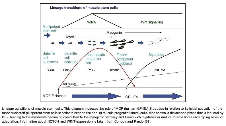
What remains, then, is what IS causing muscle growth. Right now, the likely culprit is MGF. This is a splice variant of IGF1. It would cause me a whole host of pain and stress to write more on this, as it is a somewhat cloudy and developing field with tons of supremely interesting information (and I’m at work; thankfully I work in a research lab whose focus is IGF1R), so I’ll leave you with a diagram representing how MGF and IGF1 work together to create hyperplasia:
 
W

willc86

Member
Awards
0
oh god O_O the more I know, the less I know lol!!!!
 
W

willc86

Member
Awards
0
IGF1 is thought to induce muscle hypertrophy, by distinct mechanisms. The IGF1 receptor is a tyrosine-kinase receptor which induces cellular signal transduction chains by adding phosphate groups or “phosphorylating” specific proteins within the cell. Activation of the PI3K/AKT kinases cause phosphorylation of the FOXO transcription factors, which prevents them from entering the nucleus and promoting the expression of atrophic factors, like MuRF1. The AKT pathway (often called “PKB” instead of “AKT”) also inhibits the secretion of myostatin, thereby increasing both muscle cell differentiation, and protein synthesis.(ref) Myostatin inhibition results in a positive feedback cycle, since myostatin also inhibits the AKT pathway.(ref, ref) IGF1 also activates the mTOR pathway, which is well-known to play a central role in muscle growth. Apparently, PI3K activates mTOR by moving tuberous sclerosis complexes (mTOR inhibitors) from the membrane to the cytosol.(ref) (Independent of growth factors, amino acid availability, especially leucine, regulates mTOR activity, ref.) For a more detailed discussion of the AKT pathway, see: Akt: a nexus of growth factor and cytokine signaling in determining muscle mass


what does it mean by "thereby increasing both muscle cell differentiation and protein synthesis" <-- ok I know protein synthesis, what about the other stuff
 
Torobestia

Torobestia

Well-known member
Awards
2
  • RockStar
  • Established
Myostatin is both responsible 1) for blocking satellite cells (a form of "differentiated" stem cells) from differentiating/becoming skeletal muscle cells, 2) for blocking the expression of certain skeletal muscle-specific genes in developed muscle cells, and 3) for blocking protein synthesis by inhibiting mTOR/p70s6k. So that excerpt in quotes was referring to how if AKT phosphorylation inhibits myostatin release, then by proxy it also blocks the effects of myostatin, which would lead to increased satellite cell differentiation and increased protein synthesis (it's like the foot was taken off of the brakes).
 
Thread starter Similar threads Forum Replies Date
tobias Sherman IGF-1/GH 16
J IGF-1/GH 2
S Anabolics 1
V Supplements 3
V Supplements 0

Similar threads


Top